Interface design for NT-MDT scanning probe microscope control software

Problem: Develop the user interface for the scanning probe microscope control program that’s equally fit to solve scientific, technological, theoretical, and practical problems.

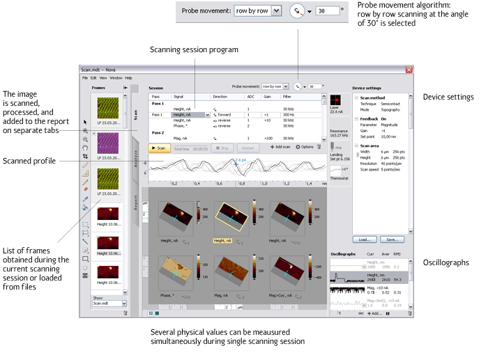
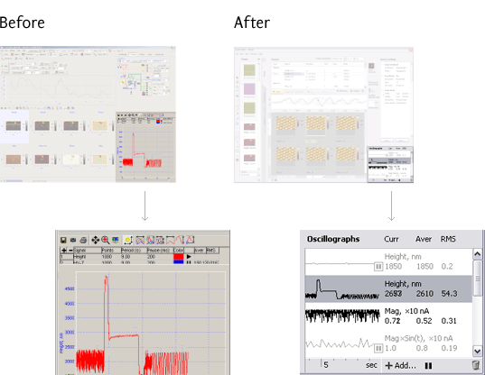
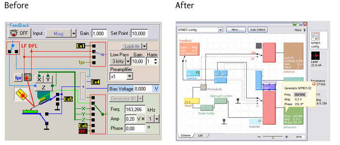
Scanning probe microscopy (SPM) is used in modern physics, chemistry, materials science, biology, medicine, and hi-tech production. Art. Lebedev Studio designed the interface of the program to control scanning probe microscopes—the program used by scientists, engineers, and technologists to adjust these complex devices and acquire SPM images in order to solve research, development, diagnostic, and quality control problems. The acquired images are further processed in a separate processing and analysis interface, which was developed by Art. Lebedev Studio as well.

Principle of scanning probe microscope operation

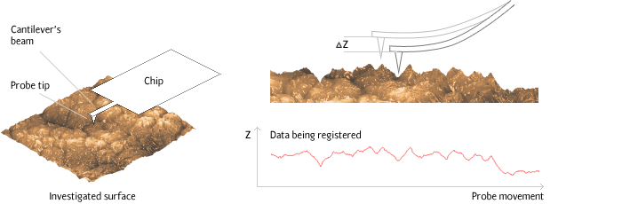
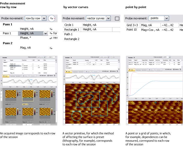
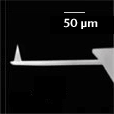
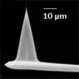
In scanning probe microscopes the micropattern of the surface and its local properties are investigated using special acerate probes. The size of the operating point of such probes is around 10 nm (0.00001 mm). The typical distance between the probe and the sample surface in scanning probe microscopes is 0.1–10 nm.

В натуральную величину С увеличением С увеличением
Probe sensor: left—about life-size; at the left-hand end of the rectangular base one can see the finest 0.1 mm-long feeler—a flexible cantilever. At the end of the cantilever, perpendicularly to its plane, there is a pointed probe invisible to the eye

The operation of scanning probe microscopes is based on different types of the probe’s contacts with the surface. In the simplest case, the point of the probe contacts the surface being investigated. The probe sensor moves along the surface, whereby the changes in the flexion of the cantilever or other values proportional to the probe’s contact to the investigated sample are registered.

Release date: March 23 2006

Cast:

artistic director
art director and interface developer
interface developer
pictographer
manager

Art. Lebedev Studio would like to thank Stanislav Leasment for his help in project implementation

All SPM images are the property of NT-MDT

Materials from the book Scanning Microscopy Basics by Mironov V.L. (Russian Academy of Sciences, the Institute of Physics of Microstructures. Nizhny Novgorod. 2004) were used

Как работает кантилевер
A simplified scheme of how SPM works: as the probe moves, the signal is read. If the probe has covered the entire stretch, the program can build a map of distribution of the values being measured and, for example, reproduce the relief of the scanned surface

Apart from simple scanning that results in a map of value distribution, there are other methods of using SPM. For example, in spectroscopy one gets a set of dependences of one value upon the other in a variety of surface points. In lithography, by affecting the sample, a certain pattern is reproduced on its surface—for example, the pattern of an electrical nanocircuit. In nanomanipulations, using the probe, one can cut a living cell or move a large molecule of a carbon nanotube to contacts prepared beforehand, thus building a nanodiode.

New interface

Work with different SPM modes and methods has been as much standardized as it was possible. For that, two concepts were set apart in the interface: a) the way the probe affects the sample and the measuring of this effect, and b) the algorithm of probe movement. This separation made possible the support of the existing methods (row-by-row scanning, spectroscopy, vector and raster lithography), their combinations, and yet-undiscovered methods. In the older version of the program the methods were realized in a linear way: a separate program was in fact developed for each of them.


Главное окно
Main window
Indicator shows the time elapsed and time remaining
The graph displays the profile of the current scanning row and the gradually dimming profiles of the previous 5-6 rows
Oscillographs allow for tracking the device’s signals in real time. In the new version the table of oscillographs takes up less space and displays several signals at the same time. If needed, the user can open the required graph in a larger separate window

The program has a full-screen mode that can be used if you don’t want to be distracted by anything while scanning, or during the presentation of the device.

The entire space can be smoothly scaled using the scrollbar in the corner. To see all images at once, the scrollbar has to be tugged to the extreme left. A double click on one of the images will enlarge it quickly. The width of the line within the round-shaped scrollbar changes during scaling

Adjustment of probe movement

Probe movement control elements are placed in the upper part of the window. They allow for selecting the type of movement (row-by-row, spiral, point-by-point, by vector curves) and determining its direction (for example, the angle of scanning for row-by-row scanning).

The direction and angle in row-by-row scanning are set using a special element that consists of four basic directions and a circular element that determines the scanning angle. The text input field can also be used for precise angle adjustment.


Scanning session

For unification of work with different SPM methods, the concept of the scanning session was introduced in the interface.

Scanning session is a program that has to be executed by the device. In the usual mode of row-by-row scanning, a list of images is preset in the session table, which need to be obtained by scanning the predetermined surface and measuring different physical values. In other modes the session can be determined in other terms:


While setting the scanning parameters, the user thus adjusts first of all the images to be obtained, not the nodes of the device.

Before: in the older interface of scanning parameters adjustment the hardware essentials were in the forefront
To edit the data in the session table, the edit-in-place method is used

Auxiliary equipment control

The indicators of the state of auxiliary equipment are so located as to allow the user to track problems with the equipment and quickly adjust the systems in smaller pop-up windows while scanning. In the older version of the program the systems were adjusted in independent full-screen windows, which took the user’s focus off the scanning process.

Indicators of the state of auxiliary equipment: laser, generator, probe, thermostats, external magnetic sources etc. After the indicator is pressed, a movable window opens, where the description of the state of the relevant node and the tools of special adjustment, for example, laser adjustment, are displayed. As long as the window is open, the indicator remains pressed

Настройка резонансной частоты генератора
Adjustment of the generator’s resonant frequency

Device configuration

A special window of the device configuration allows for adding new hardware components to the system in a visual mode.


The earlier scheme of the device used to be preset, so the user could only adjust some of the device’s parameters. The new scheme reflects the real device configuration and makes possible the adjustment of all its components

The components can be connected to each other either directly or through switch boxes. The switch boxes allow for switching links between components with one click.

Коммутатор
The switched-on links between the components are displayed as a continuous black line, switched-off links—as a grey dashed line. A port to which nothing is connected is displayed as an empty square
Components are searched for by name and parameters simultaneously with the input

Project

In 2004 Art. Lebedev Studio started working on the user interface of software products developed by NT-MDT for controlling probe microscopes and processing the data acquired using them.

The interface of the probe microscope control program was developed in February–December 2005.

The image of our logo that was acquired using the method of raster lithography on a 1×0.3 micron titanium film. The width of one bar of the bar code is about one hundred atoms

Order a design...