|
To increase the precision of diagnostics, the doctor can use different diagrams built in two or three dimensions based on the parameters of the original X-ray. In one of the modes a two-dimensional X-ray is displayed as a three-dimensional surface whose third coordinate is brightness. For the visualization of the three-dimensional form, a special color scale (pseudospectrum) coupled with imitated lighting were used. |
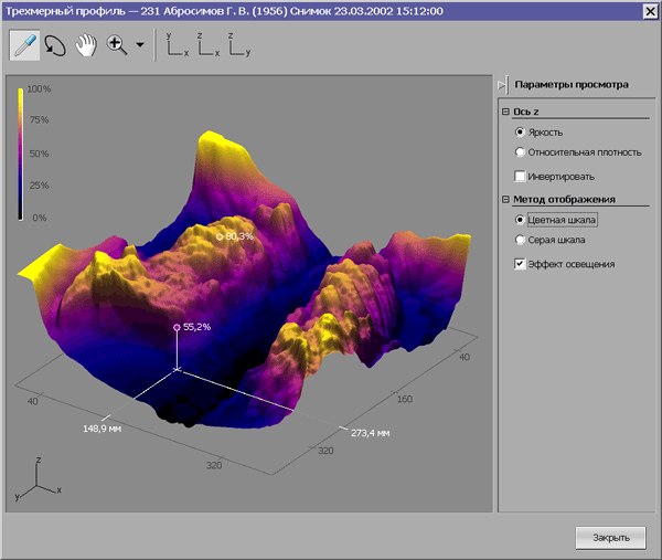 Pseudospectral color coding helps the doctor immediately pinpoint the areas with the same brightness
|
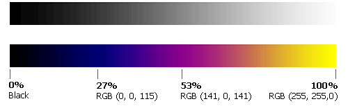 Black-and-white scale and pseudospectrum
The color scale was selected based on the following two requirements:
|
 In medical devices and programs a physical spectrum is often used, although it’s totally irrelevant for the visualization of the smooth changes in values |
|
To scale all two-dimensional graphs, Art. Lebedev Studio specialists devised a special control element. The graph is displayed at two windows at a time: in a small window the entire range of values is shown, while the big one shows an enlarged fragment of the graph. |
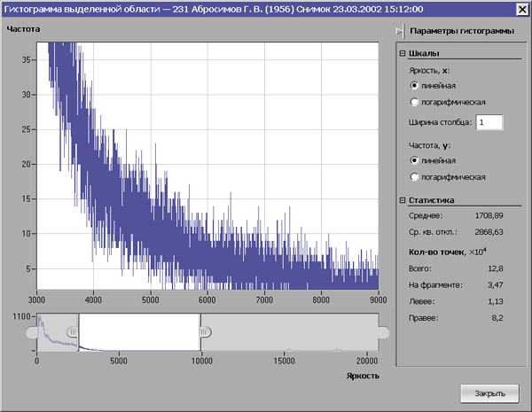 With the help of two sliders, the user can choose on the X-axis the range he is interested in, and the optimal scale of the graph will be selected automatically
|
|
Even the bar chart interface that’s customary for many computer images processing software was substantially improved: the color of the selected areas of the range is now different from that of the rest of the range; the fields for entering limit values and black and white pipettes are located under the relevant edges of the black-and-white scale. |
 |
